1 min read 0

Case of the Week: Odontogenic Myxoma

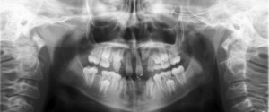
This week is another case and video.  This case shows a well-localized radiolucent area between the mandibular left first premolar and canine on the reconstructed pantomograph. It is causing displacement of the roots of these teeth. The axial and sagittal views show thinning with displacement of the facial cortical plate…
1 min read 0

Case of the Week: Ectodermal Dysplasia

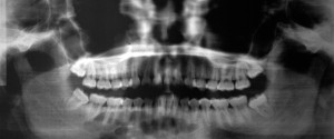
This week’s case is opposite of a case from a few weeks ago (Cleidocranial Dysplasia) in that it presents with absence of teeth (oligodontia). In some extreme cases, no teeth may develop at all (referred to as anodontia). The teeth that are present typically have a conical shaped crown. Here…
1 min read 2

Case of the Week: Hyperparathyroidism with a Brown Tumor

This week I have a case with two interesting findings. The first is hyperparathyroidism which typically presents with a ground-glass appearance in the jaws instead of normal bone trabeculation.  This appearance is visible in all bones of the body and not just the jaws.  In some cases (~10%) there is…
3 min read 0

Locate the Object: May 2014 ANSWER

Here is the answer for the May 2014 Locate the Object. I will be going over both image shift and SLOB (Same-Lingual, Opposite-Buccal). . Image shift Before starting to use the image shift principle it is important to know/remember two key points Images move in the opposite direction from the…