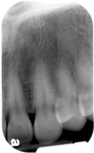
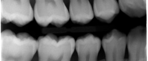
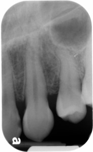
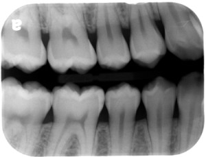
Here are the carious lesions for the 4 radiographs posted earlier this week.
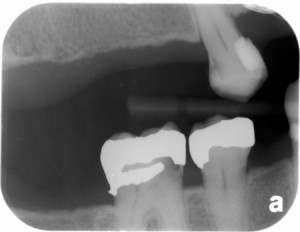
1.
- Maxillary left lateral incisor (#10) distal
- Maxillary left canine (#11) mesial
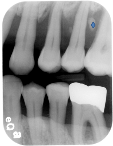
- Mandibular right second premolar (#29) distal
- Mandibular right first molar (#30) mesial
- Maxillary right first premolar (#5) distal
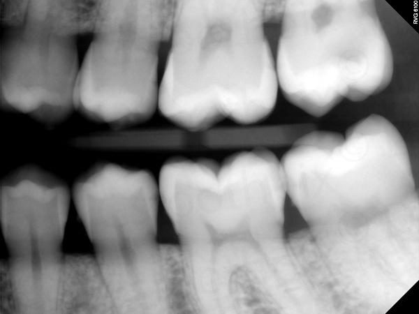
- Mandibular right first molar (#30) mesial
- Maxillary left central incisor (#9) distal possible
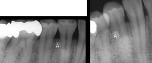
- Maxillary left lateral incisor (#10) mesial and distal
 If you have any questions or comments, please leave them below. Thanks and enjoy!
Hi Dr. G,
On third picture, is there caries on the distal of mandibular right canine?
Is the opened contact between the premolar and molar on this radiograph still considered a regular openned contact “created” by the bitewing?
What I mean is, when we should consider that the contact is open looking on bitewings. Or we can’t say just by radiographs? 😀
On the third picture, there is a cervical composite restoration and cervical burnout apical to this but no caries.
As for when a contact is open on bitewing radiographs, there is no overlap of the adjacent teeth. This does not mean there is a true open contact between two adjacent teeth meaning they are not touching. Is this what you are asking?
Now I see the composite on the canine. I would had mark this as caries on the exam and would lose the question. Thanks for showing me the difference.
So when can we say that there is an open contact just looking on a bitewing radiograph? Is there a parameter for that? E.g. space bigger than 1mm we may consider as an open contact.
The only way to tell if adjacent teeth are not touching is to look for a visible radiolucent line between the teeth indicating air between the two teeth. It’s a little tricky in some cases. If you have digital images you can lighten the image to see if the radiolucent area between the two teeth stays the same radiolucency as above and below the contact. Does this help?
It helped a lot! 🙂
I didn’t know that technique with the digital images.
Thanks again, Dr. G!