Anatomy Monday: Gubernaculum Dentis 2


This week I have a case of anatomy that I more commonly see on CBCT images but have occasionally come across on 2D radiographs; the gubernaculum dentis. The gubernaculum dentis is a canal of connective tissue from a developing tooth bud to the overlying mucosa. It presents as a radiolucent band from the developing tooth bud to the crest of the alveolar ridge.
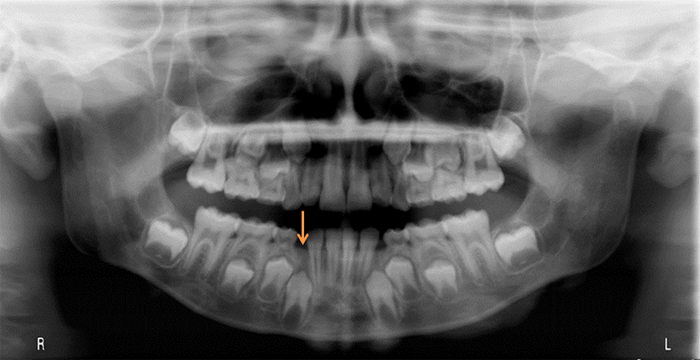
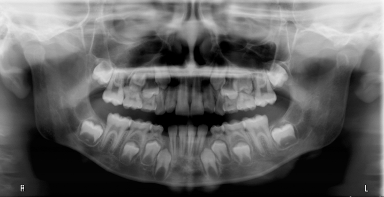
Pantomograph - orange arrow noting the gubernaculum dentis from the developing permanent canine.

Pantomograph – orange arrow noting the gubernaculum dentis from the developing permanent canine.

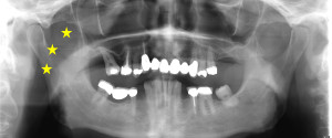
Pantomograph - gubernaculum dentis as a vertical radiolucent band superior to the crown of the developing right mandibular canine.

Pantomograph – gubernaculum dentis as a vertical radiolucent band superior to the crown of the developing right mandibular canine.

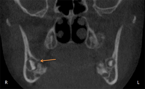
CBCT coronal view - orange arrow noting the gubernaculum dentis

CBCT coronal view – orange arrow noting the gubernaculum dentis

CBCT coronal view - the gubernaculum dentis as a horizontal radiolucent band from the developing right mandibular molar to the crest of the alveolar ridge.
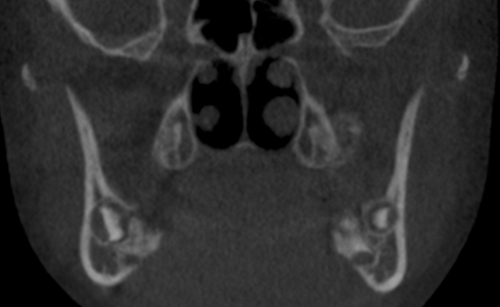
CBCT coronal view – the gubernaculum dentis as a horizontal radiolucent band from the developing right mandibular molar to the crest of the alveolar ridge.

If you have any questions or comments about the gubernaculum dentis, please leave them below. Thanks and enjoy!

2 thoughts on “Anatomy Monday: Gubernaculum Dentis

Comments are closed.