1 min read 4

Educational Video: Attrition

This week I have a case of attrition along with an educational video.  First the case; this is an example of attrition on the lingual surfaces of the maxillary incisors.  It presents with the crown having an increased radiolucent appearance and a defined horizontal line near the cemento-enamel junction where…
1 min read 4

Anatomy Monday: Posterior Superior Alveolar Canal

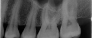
This last canal is one that is always fun to see and a great quiz question for students (at least for me but then again I am a radiology nerd :D).  The posterior superior alveolar canal is most commonly seen on posterior maxillary periapical radiographs. It presents as a thin…
1 min read 0

Case of the Week: Talon Cusp

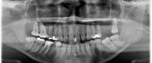
This week I have a case of a talon cusp so large it’s evident on a pantomograph.  While there is more than one talon cusp evident on this radiograph, it’s the one on the maxillary left lateral incisor that is nearly as big as the crown that’s amazing :).  Note…
3 min read 0

Locate the Object: July 2013 Answers

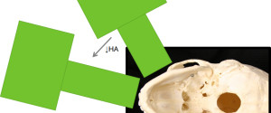
Now onto the answers for the July 2013 Locate the Object.  I will be going over both image shift and SLOB (Same-Lingual, Opposite-Buccal) to determine the location of the pin superimposed over the first premolar. Image shift Before starting to use the image shift principle it is important to know…