1 min read 0

Case of the Week: Pulp Stones

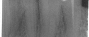
This week is a case of two elongated pulp stones along with an educational video. The case shows an oblong radiopaque mass in the pulp chamber and root canal space of the canine and first premolar. Note how the chamber and canal space is enlarged around the radiopaque mass. Here’s…
1 min read 0

Educational Video: Sinusitis

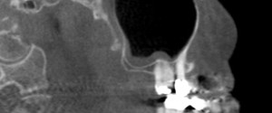
This week I have a case of sinusitis on cone beam CT along with another educational video. 🙂 On the cone beam CT images note the radiopaque band that follows the floor and the posterior border on the sagittal view and the floor and medial border of the left maxillary…
1 min read 3

Locate the Object: August 2013

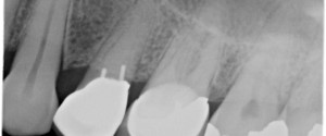
Onto another fun case to use your image shift and SLOB (Same-Lingual, Opposite-Buccal) information. The object in question is the left lateral incisor. Where is it (buccal or lingual) in relation to the left central incisor? The answer will be coming in two weeks (August 27th).  You can answer either…
1 min read 0

Anatomy Monday: Anterior Superior Alveolar Canal (???)

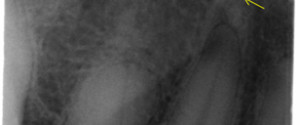
I’m going to start out this week’s canal as a challenge. I’ve come across two cases recently with canals that appear to be heading towards the maxillary canine. Based on my research and review of anatomy I concluded this was possibly the anterior superior alveolar canal.  After discussing this case…
1 min read 0

Case of the Week: Palatal Tori

This week I have a case of palatal tori evident not only on a periapical radiograph but a pantomograph as well.  On the periapical radiograph it appears as a radiopaque mass superior to the maxillary teeth. The pantomograph shows a well-defined horizontal radiopaque band directly inferior to the floor of…