1 min read 0

Anatomy Monday: Nasolacrimal Canal

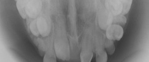
The nasolacrimal duct is most commonly seen on occlusal radiographs specifically the anterior maxillary occlusal radiograph and standard maxillary occlusal radiograph.  It presents as an ovoid radiolucent area with a thin radiopaque border at the lateral edge of the nasal cavity in the region of the molars. If you have…
1 min read 0

Case of the Week: Hypercementosis

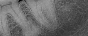
This week is a fun case of hypercementosis at the apex of a molar root.  Note on the distal root of the first molar a radiopaque mass with a thin radiolucent line around it. This shows that the mass is attached to the root surface with the periodontal ligament space…
1 min read 0

Anatomy Monday: Infraorbital Canal

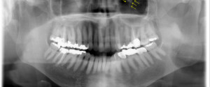
The infraorbital canal is seen on extraoral radiographs specifically pantomographs.  It appears as a diagonal radiolucent band with two thin radiopaque borders superimposed over the inferior border of the orbital rim. If you have any questions or comments about the infraorbital canal and its appearance on radiographs, please leave them…
1 min read 0

Case of the Week: Enamel Pearls

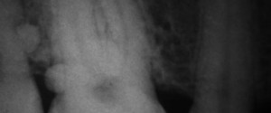
I have to send a big thank you out to Dr. Marc I Semper for sharing this case of enamel pearls with me.  He was also kind enough to share a link to a great overview page about enamel pearls by Marco A Versiani.  Below I’ll let you find the…