This last canal is one that is always fun to see and a great quiz question for students (at least for me but then again I am a radiology nerd :D). The posterior superior alveolar canal is most commonly seen on posterior maxillary periapical radiographs. It presents as a thin curved radiolucent line coursing horizontally through the maxillary sinus. The canal is actually in the lateral border of the maxillary sinus but appears to be within the sinus on 2D radiographs.
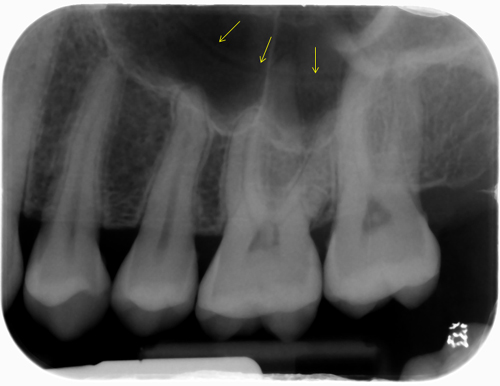
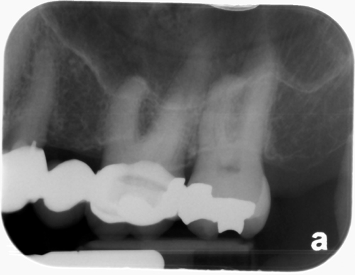 If you have any questions or comments about the posterior superior alveolar canal and its appearance on radiographs, please leave them below. Thanks and enjoy!
Superb! I’ll check my cbct’s for this finding. Cheers, Marc
Great! If you find some fun ones to share please send them my way. 🙂
Can this PSA canal appear as a wave of radiolucent band instead of a thin curved radiolucent line?
I haven’t come across one with a wave pattern. I’d be curious to see what exactly it is you are referring to.