1 min read 0

Anatomy Monday: Infraorbital Canal

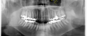
The infraorbital canal is seen on extraoral radiographs specifically pantomographs.  It appears as a diagonal radiolucent band with two thin radiopaque borders superimposed over the inferior border of the orbital rim. If you have any questions or comments about the infraorbital canal and its appearance on radiographs, please leave them…
1 min read 0

Case of the Week: Enamel Pearls

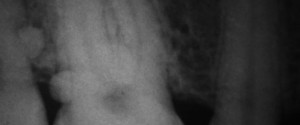
I have to send a big thank you out to Dr. Marc I Semper for sharing this case of enamel pearls with me.  He was also kind enough to share a link to a great overview page about enamel pearls by Marco A Versiani.  Below I’ll let you find the…
1 min read 1

Locate the Object: July 2013

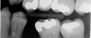
Being that image shift and the SLOB rule (Same-Lingual, Opposite-Buccal) are topics I am frequently asked about and popular viewed pages on this site, I decided to start posting practice cases once a month for you to put that knowledge to work. This first case has three radiographs to use.…
1 min read 0

Anatomy Monday: Nasopalatine Canal

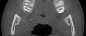
It’s time for the next canal: the nasopalatine canal.  This canal may also be referred to as the incisive canal.  It is seen on both intraoral radiographs and extraoral radiographs.  The nasopalatine canal presents as a vertical radiolucent band between the roots of the maxillary central incisors superiorly to the…