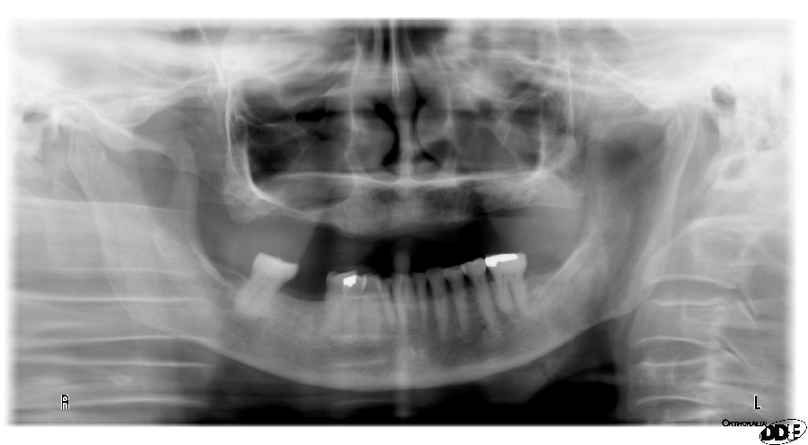
This week is another soft tissue entity that can be seen on both intraoral and extraoral radiographs. The nasolabial fold presents as a diagonal transition line. A transition line is seen as a defined line where part of the radiograph appears more radiopaque due to superimposition of soft tissue. The nasolabial fold is seen over the maxillary premolar and canine regions.
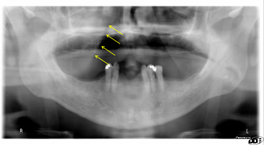 Yellow arrows noting diagonal transition line of nasolabial fold
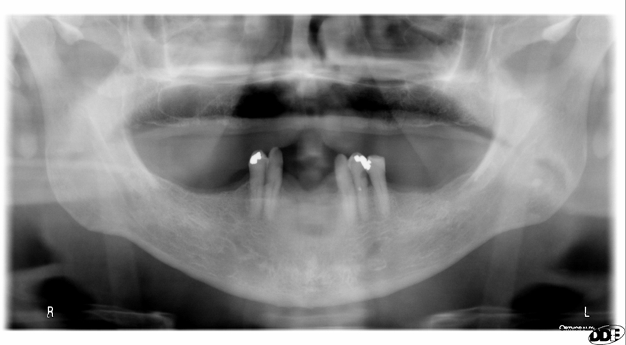 Pantomograph without arrows. Nasolabial fold noted on both right and left sides
Periapical Radiographs
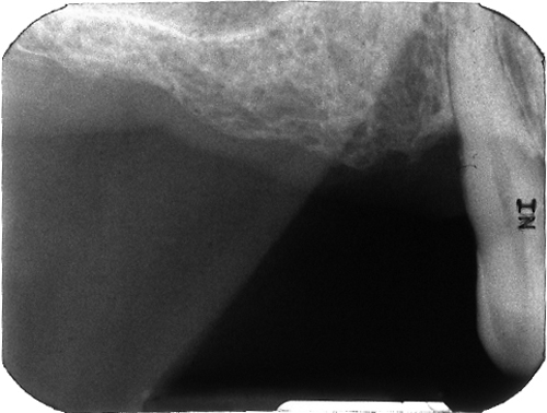
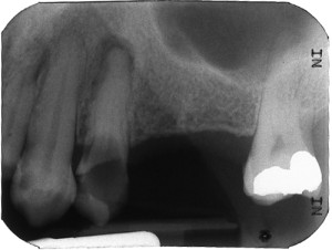 If you have any questions or comments about the nasolabial fold appearance on radiographs, please leave it in the comments below. Thanks and enjoy!


Does nasolabial fold landmark determine the anterior and posterior regions ?
Not necessarily as it can appear as far posterior as the first molar and anterior as the canine. So it’s not a hard separation between the two.