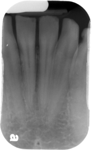
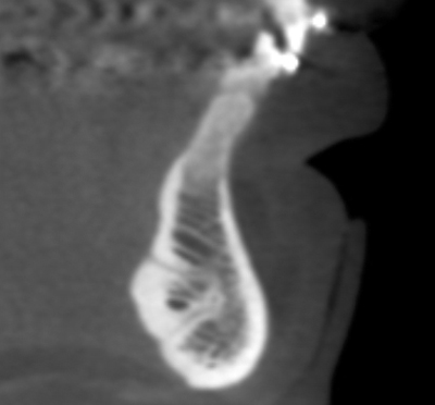
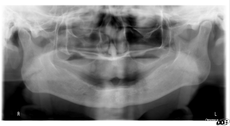
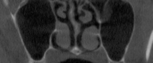
It’s another foramen in the mandible. While there are multiple lingual foramina in the mandible, the most commonly seen one is in the anterior mandible on the midline. It appears as a round radiolucent entity with a thin to thick corticated border. It may be single or multiple (up to 3 however this is more commonly seen on CBCT). Check out some examples below.
Periapical Radiograph
CBCT sagittal view showing two lingual foramina

2 thoughts on “Anatomy Monday: Lingual Foramen (mandible)”
Comments are closed.