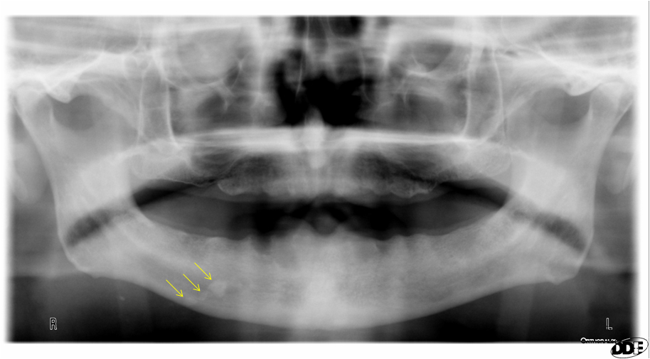
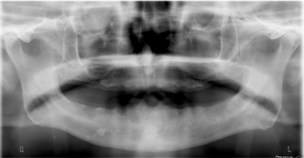
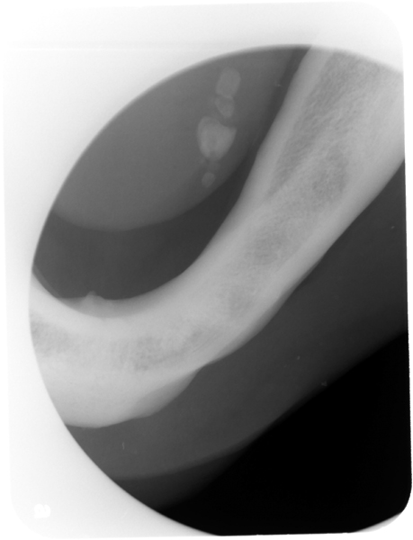
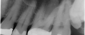
This week I have a neat example of a sialolith on a pantomograph. This was an incidental finding at a new patient exam. Sialoliths tend to be single, but this case has multiple calcifications within the duct of the submandibular salivary gland. On the pantomograph there are multiple, irregular shaped radiopaque masses over the inferior portion of the right mandible (first image has arrows). The patient then gave a history of frequent swelling episodes in the floor of their mouth with popping noises coming out of their mouth when they would press on the swelling. A mandibular true occlusal was made to verify the presence of a sialolith. On the mandibular true occlusal radiograph, you can see the multiple irregular shaped masses that are located in the floor of the mouth.
For more information and other radiographs of sialoliths check out the page on sialoliths.
Enjoy!
