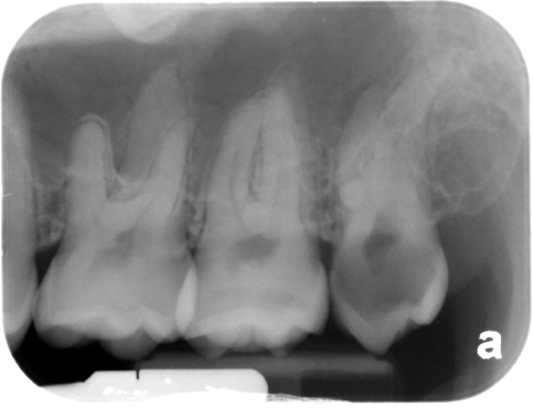
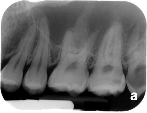
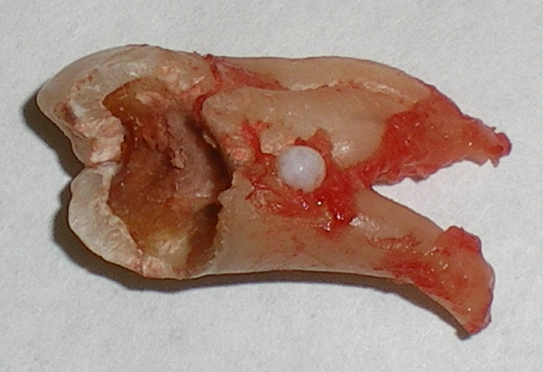
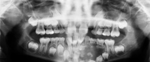
This week I am showing a case of an enamel pearl along with the clinical photo of the tooth after it was extracted. Enamel pearls are an extra ‘blob’ of enamel near the furcation area on molars. They will present as circular radiopaque areas near the furcation. They can be tricky to interpret on radiographs due to superimposition of adjacent roots on some periapicals and bitewings. For this reason it is important to have at least two radiographs with different angulations (either vertical or horizontal). This will show if the enamel pearl is seen on both radiographs or if the increased radiopaque area was due to superimposition of adjacent roots. This case was an enamel pearl on a maxillary third molar. Note the circular radiopaque area near the furcation. The clinical photo was at the time of the extraction showing the enamel pearl on the tooth. (This is the first case on the enamel pearl page.)
For more information and other radiographs of enamel pearls check out the page on enamel pearls.
Enjoy!
