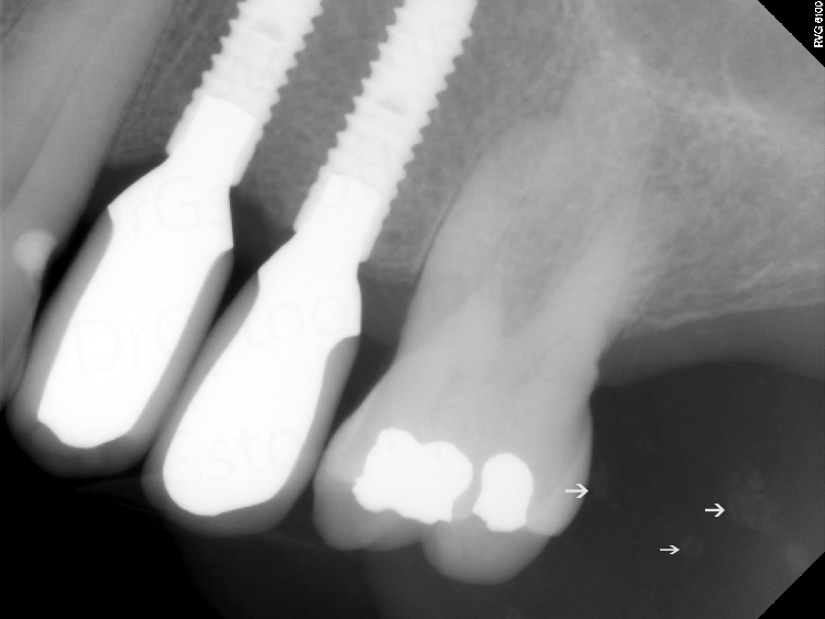
This week I have an example of a very large radicular cyst (sometimes referred to as a periapical cyst) that was encroaching on the maxillary sinus. The patient presented with no symptoms during a new patient exam. A pantomograph was made. On the pantomograph, a well-defined ovoid radiolucent entity is seen superior to the maxillary right premolars and canine. A periapical showed lose of bone apically to these teeth with gross decay of the premolars. Vitality tests showed the premolars were non-vital. Due to the proximity and effect on the maxillary sinus a cone beam CT was ordered. Below are the images showing the radicular cyst and it’s effect on the maxillary sinus.


CBCT images – coronal and sagittal views


For more information and other radiographs of radicular cysts check out the page on radicular cyst.
Enjoy!