cervical spine


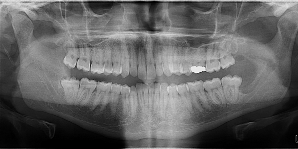
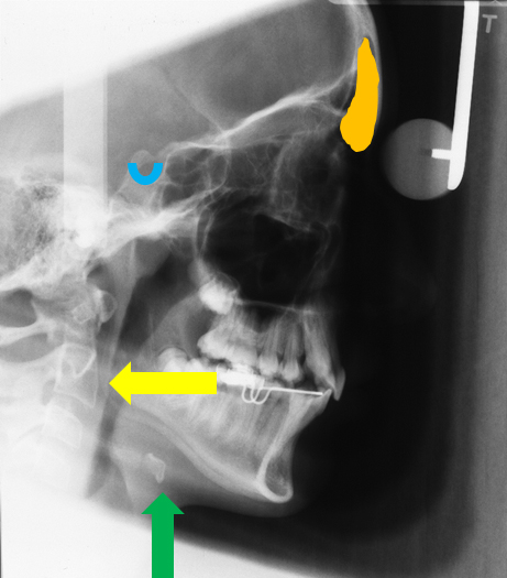
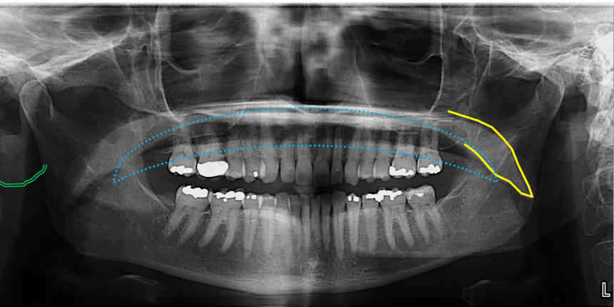
Case of the week: Normal pantomograph imaging mimicking disease 4

This week I wanted to show an area on pantomographs that I am frequently asked about.  The area I am talking about is the radiolucent area as noted by the yellow areas inferior to the mandibular posterior teeth. This area is typically where an observers eye are drawn due to […]


Anatomy on Radiographs: Extraoral Radiographs (Lateral Cephalometric Skull) 2

This week is extraoral radiographs, specifically the lateral cephalometric skull radiograph.  This will be a short and sweet post only going over basic anatomy. I am not covering most of the anatomical landmarks used in orthodontics, but if you would like a post on this, please let me know and […]


Anatomy on Radiographs: Pantomographs Part 2 10

Now onto other structures visible on a pantomograph. Midface The orbital rim (green dots) appears as a thick semi –circular radiopaque band superior to the maxillary sinuses. The pterygomaxillary fissure (blue area) appears as an inverted teardrop shaped radiolucent area with a thin radiopaque border. It is directly lateral to […]