
Here is the answer for the May 2014 Locate the Object. I will be going over both image shift and SLOB (Same-Lingual, Opposite-Buccal). .
Image shift
Before starting to use the image shift principle it is important to know/remember two key points
- Images move in the opposite direction from the movement of the source.
- Images of objects farther from the image receptor will move more (aka objects (images) more facial/buccal will appear to move more).
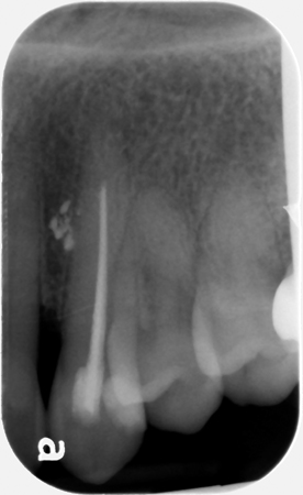
 The first thing to do is pick a stationary object that is seen on both radiographs and appears to move in comparison to the extruded sealer/gutta percha. On these radiographs, I will use the gutta percha in the root canal space of the canine. The most obvious angle change is to the horizontal angle. Starting with the canine periapical radiograph and moving to the lateral incisor periapical radiograph, the source of radiation (tubehead) moves anteriorly decreasing the horizontal angle. According to point 1 above, this means the images move posterior.
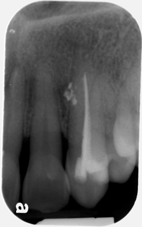
Looking at the second radiograph (lateral incisor periapical radiograph), we need to compare the image movement of the extruded sealer/gutta percha versus the gutta percha in the root canal space of the canine to see which object moved more posterior following point 2 listed above.
The extruded sealer/gutta percha appears to be more posterior on the radiograph meaning it is farther from the image receptor compared to the gutta percha in the root canal space of the canine. This gives us an answer of the extruded sealer/gutta percha being to the buccal/facial of the gutta percha in the root canal space of the canine (hence to buccal/facial of the canine root).
SLOB (Same-Lingual, Opposite-Buccal)
The first thing to do is again pick an object with a known fixed location – the gutta percha in the root canal space of the canine.
The second thing to do is determine which direction we are moving from the canine periapical radiograph to the lateral incisor periapical radiograph and the answer would be – mesial.
Lastly, on the lateral incisor periapical radiograph determine what direction the extruded sealer/gutta percha appears to have moved in relation to the gutta percha in the root canal space of the canine – distal. Here is where the acronym comes into play. Did the unknown object move in the SAME direction as the radiographs or in the OPPOSITE direction? Our answer is – opposite and the acronym states that opposite is buccal, so the extruded sealer/gutta percha is to the buccal of the gutta percha in the root canal space of the canine (hence buccal to the root).
Another case will be coming next month. If you have any questions or comments or would like two cases a month, please let me know below. Thanks and enjoy!