
Last week I showed the superior foramina of the nasopalatine canal and this week is the inferior foramen; the incisive foramen. The incisive foramen presents as a round to ovoid radiolucent entity between the maxillary central incisors. When the width of the incisive foramen is 10 mm (1 cm) or larger a nasopalatine canal cyst should be considered.
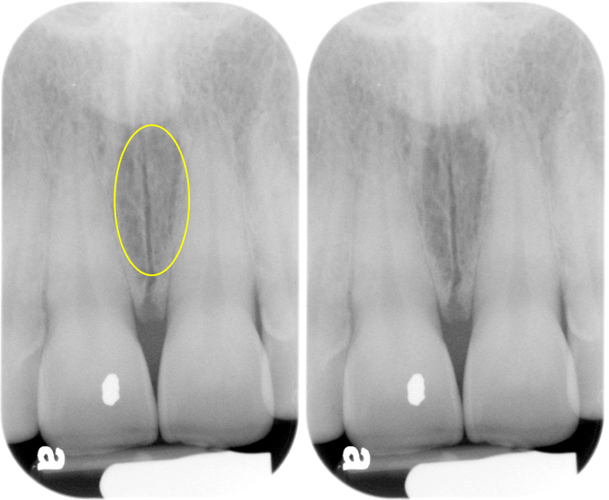
Maxillary central incisors periapical radiograph.
Left – yellow circle showing the incisive foramen.
Right – incisive foramen as an ovoid radiolucent entity between the maxillary central incisors.
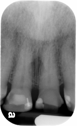
Maxillary central incisors periapical radiograph showing the incisive foramen as a round radiolucent entity between the maxillary central incisors.
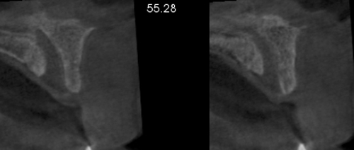
CBCT cross-sectional slices showing the inferior opening of the nasopalatine canal as a discontinuity of the inferior aspect of the maxilla.
If you have any questions or comments about the incisive foramen, please leave them in the comments below. Thanks and enjoy!