The entire acronym of DEBT does not apply to both digital radiography (DR) and computed radiography (CR) systems. Since both systems are a little different with artifacts I have broken them up into two separate posts.
Increased density
- E (increased exposure to x rays)
E =↑ Exposure (x rays)
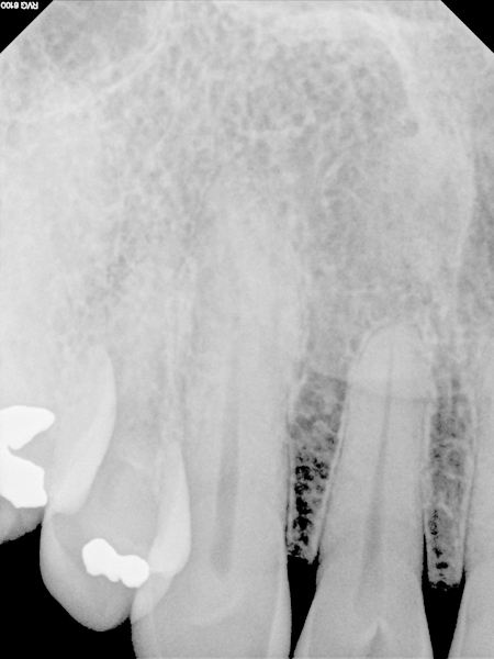
This occurs when the sensor is overexposed (i.e. using a molar setting for an anterior radiograph).
Overall increased density due to incorrect time setting.
Decreased density
- E (decreased exposure to x rays)
E = ↓Exposure (x rays)
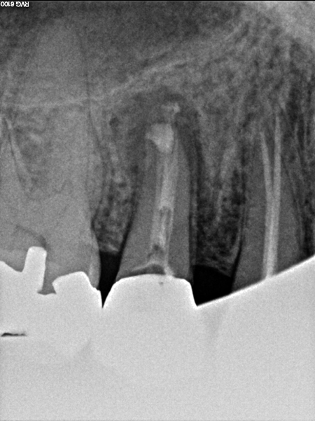
This occurs when the sensor is underexposed or not exposed at all. There are two ways this can occur. The first is decreased time of radiographic exposure (i.e. using a anterior time setting for a molar radiograph) creating an overall whiter image.
Overall decreased density due to incorrect time setting.
The second is by incorrect alignment with the PID/cone such that the entire sensor is not exposed to x rays. This will result in a white area of the final image where no x rays exposed the sensor. This white area may have a straight edge (rectangular PID) or a curved edge (round PID).
Round cone cut.
Next = Computed Radiography (CR) artifacts
If you have any questions, please let me know.
Thanks and enjoy!


