Today is covering analog/film artifacts that are of decreased density of the final image. Note: when referring to artifacts, the correct term is density and not radiolucent/radiopaque. The latter terms are used when referring to a part of the patient seen on a radiograph.
D = ↓Development
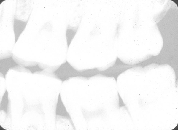
This occurs when there a decreased development of the radiographic film meaning less time the film is in contact with developer. This will appears as an overall whiter image.
Decreased overall development.
E = ↓Exposure
This occurs when the radiographic film is underexposed or not exposed at all. There are three ways this can occur. The first is decreased time of radiographic exposure (i.e. using a anterior time setting for a molar radiograph) creating an overall whiter image.
Overall decreased density due to incorrect time setting.
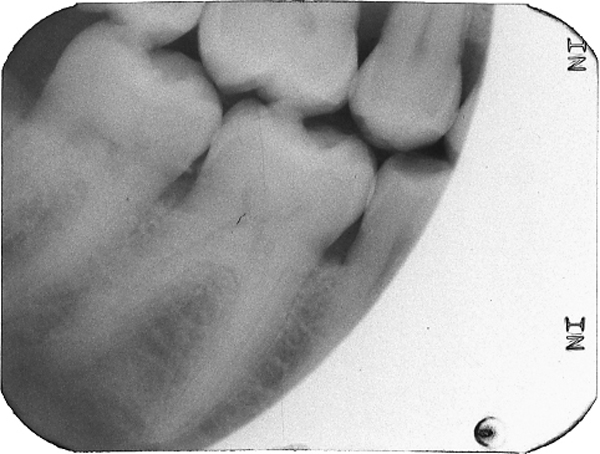
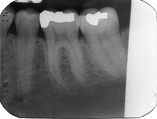
The second is by incorrect alignment with the PID/cone such that the entire film is not exposed to x rays. This will result in a white area of the final image where no x rays exposed the film. This white area may have a straight edge (rectangular PID) or a curved edge (round PID).
Round cone cut.
Rectangular cone cut.
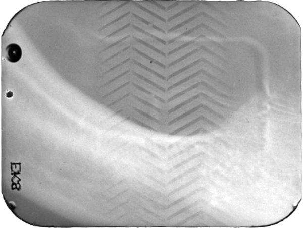
The last is by placing the film in the patients mouth backwards and exposing through the lead foil. The lead foil will stop some of the x rays resulting in an underexposure of the film. The pattern of the lead foil will be visible on the final image.
Film placed backwards in patient mouth showing pattern on lead foil.
B = No Bend
This is pretty straight forward and it means that there is no bend of the film. This does not result in a whiter area of the image and therefore doesn’t apply.
T = ↓Temperature (of the developer)
This last letter is again closely tied with the first letter as it relates to the temperature of the developer. If the temperature of the developer is decreased, this results in an overall decreased development of the film. This presents as an overall whiter image.
Decreased temperature of developer creating an overall blacker image.
Next = Digital (DR and CR) and artifacts
Thanks to Dr. John Brand for some of these images of artifacts.
I realize some of these artifacts have very similar appearances, hence I used the same images. The idea is to be aware of all the possible causes of an artifact such that it can be corrected if the radiograph must be remade. If you have any questions, please let me know.
Thanks and enjoy!


